Mode
Text Size
Log in / Sign up

AI-driven and traditional imaging markers provide a structured framework for predicting hematoma expansion in acute ICHAI tools may better predict bleeding in the brain

AI-generated summary of the cited source, checked by automated accuracy review. How we work

Key Takeaway
Note that both traditional markers and AI-driven methods provide a framework for predicting hematoma expansion in acute ICH.

This systematic review examines the evolution of imaging-based prediction methods for hematoma expansion (HE), revised HE (rHE), and ultra-early hematoma growth (uHG) in patients with acute intracerebral hemorrhage. The scope includes a comparison between traditional markers, such as CTA spot sign and NCCT signs, and modern AI-driven methodologies including radiomics, deep learning, and multi-task learning.

The review synthesizes the transition from established radiological indicators to advanced computational models. While specific predictive values for individual trials were not reported, the synthesis provides a structured framework to guide clinical practice regarding how these tools can identify patients at risk of hematoma expansion.

Authors note several limitations regarding current technologies, specifically concerning model interpretability, generalizability, and translational gaps. These factors may impact the immediate integration of AI-driven models into routine clinical workflows. The review serves as a foundational summary for clinicians to understand the current landscape of predictive tools in acute ICH management.

How this fits prior evidence

This systematic review addresses a gap in the existing evidence by providing a structured framework for predicting hematoma expansion using both traditional and AI-based methods. It complements the previously reported integrative monitoring framework for intracerebral hemorrhage, which suggested that current monitoring may miss dynamic injury. While previous findings focused on surgical interventions like MIPS or ES and medical management such as triple pill therapy, this review focuses specifically on the predictive imaging tools available for acute ICH management.

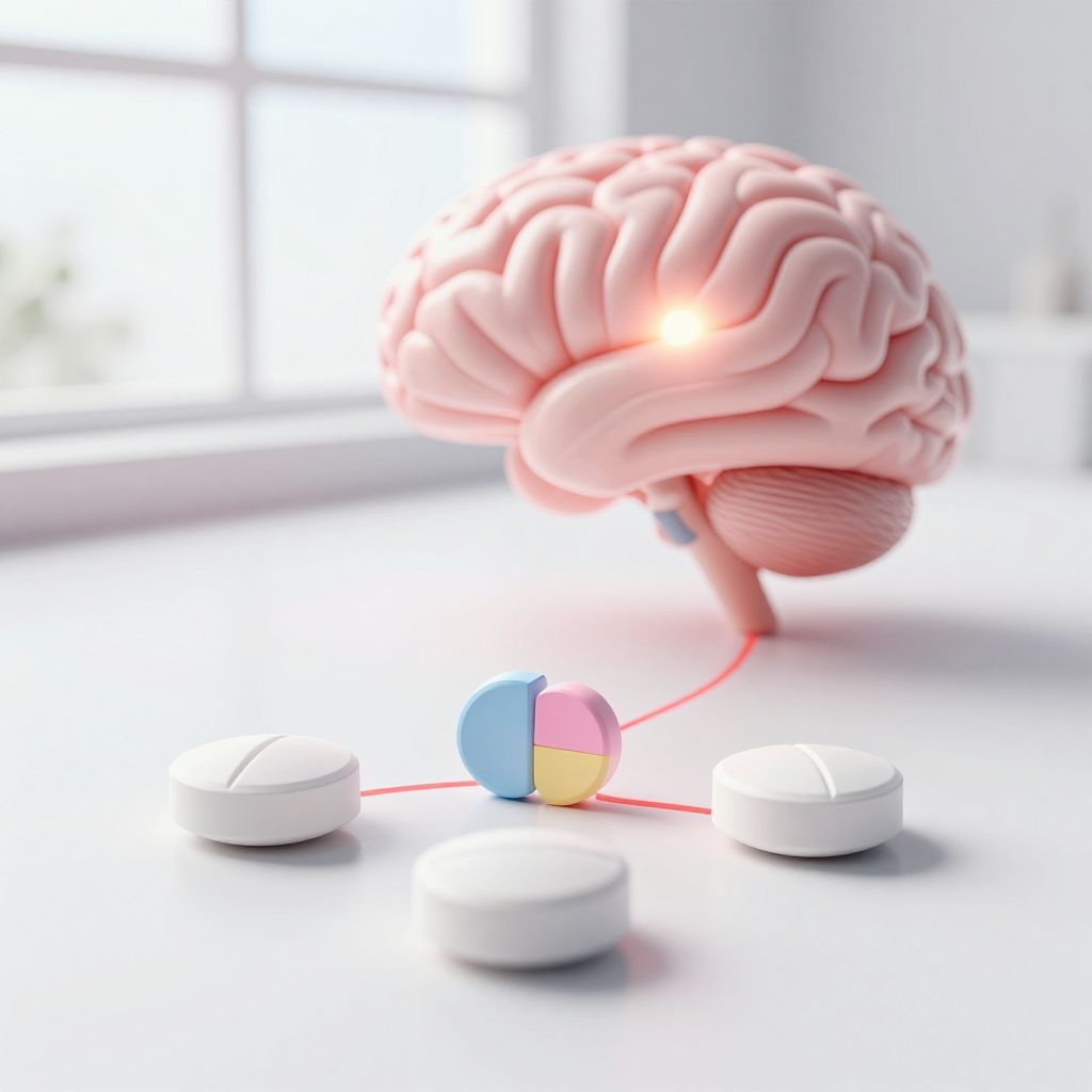
When a person has a bleeding stroke (intracerebral hemorrhage), the biggest fear is that the bleed will grow. Doctors use CT scans to spot early warning signs, but those signs are not always reliable. A new review of the research suggests that artificial intelligence tools may do a better job at predicting which bleeds will expand.

The review looked at studies comparing traditional imaging markers, like the spot sign on a CT angiogram, with newer AI methods such as radiomics and deep learning. AI can pick up on subtle patterns in scans that the human eye might miss. The hope is that better prediction could help doctors decide who needs more aggressive treatment.

But the review also points out important caveats. Many of the AI models are complex and hard for doctors to interpret. They have not been tested widely in different hospitals or patient groups. And there is still a gap between developing these tools and using them in real-world care. So while the potential is real, we are not there yet.

What this means for you:
AI may improve prediction of brain bleed growth, but more work is needed before it helps patients.

Common questions

What is hematoma expansion?

Hematoma expansion is when a bleed in the brain gets larger after the first scan. It is a major concern in patients with intracerebral hemorrhage because it can lead to worse outcomes.

How do doctors currently predict hematoma expansion?

Doctors use CT scans to look for signs like the spot sign on a CT angiogram or certain patterns on a non-contrast CT. These traditional markers help estimate the risk, but they are not always accurate.

What AI methods are being studied for this?

Researchers are testing AI methods like radiomics, deep learning, and multi-task learning. These tools analyze scan images in more detail than the human eye, potentially finding patterns that predict expansion better.

Are these AI tools ready for use in hospitals?

Not yet. The review notes that many AI models are hard for doctors to interpret, have not been tested in diverse settings, and there is a gap between research and real-world use. More work is needed before they can be widely adopted.

Study Details

Study typeSystematic review
EvidenceLevel 1
PublishedJun 2026
View Original Abstract ↓
Hematoma expansion (HE) is a critical and modifiable event following acute intracerebral hemorrhage (ICH). Predicting HE accurately can inform individualized treatment and improve patient outcomes. This review systematically outlines the evolution of imaging-based HE prediction. We first define the core concepts of traditional HE, revised HE (rHE), and ultra-early hematoma growth (uHG). We then summarize predictive studies that employ traditional imaging markers, such as the computed tomography angiography (CTA) spot sign, non-contrast CT (NCCT) signs, and combined clinical-imaging scoring systems. Subsequent sections focus on AI-driven methodologies, encompassing radiomics, deep learning, and multi-task learning. The discussion extends to precision prediction through multimodal data fusion and subgroup analyses based on hemorrhage location and onset time. Finally, we address persistent challenges, including model interpretability, generalizability, and translational gaps, and suggest future directions involving federated learning, explainable AI, dynamic prediction, and closed-loop decision systems. This review offers a structured framework to guide both clinical practice and future research.
Free Newsletter

Clinical research that matters. Delivered to your inbox.

Join thousands of clinicians and researchers. No spam, unsubscribe anytime.