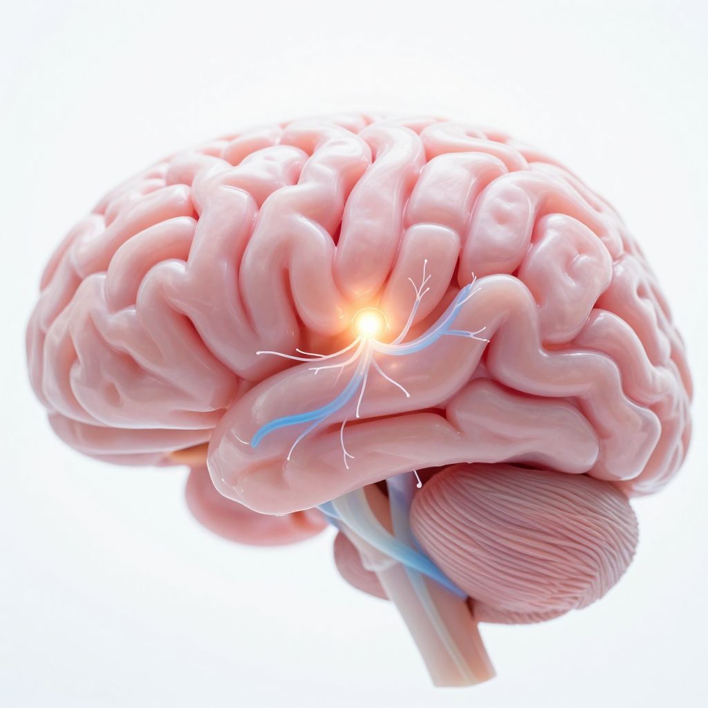
If you could see depression on a brain scan, what would it look like? Researchers tried to answer this by analyzing 42 previous studies that scanned a specific brain area—the subgenual anterior cingulate cortex (sgACC), which is involved in emotion—in people with major depression who weren't on medication. They compared these scans to those of healthy people. The results were anything but clear-cut. For brain structure, half the studies found this area was smaller in people with depression, while the other half found no difference at all. For brain activity during tasks, some studies found it was overactive, others found no change. The most consistent finding was that this emotional hub seemed less connected to other parts of the brain when at rest. It's crucial to understand that these are just associations from pictures of the brain; they don't tell us if these differences cause depression or are a result of it. The mixed results highlight how complex depression is in the brain and why a single, simple biomarker has been so hard to find.
Meta-analysis finds mixed MRI patterns in subgenual cingulate cortex in medication-free MDD patientsWhat does depression look like in the brain? The picture is complicated
AI-generated summary of the cited source, checked by automated accuracy review. How we work
This systematic review and meta-analysis synthesized evidence from 42 publications comparing medication-free patients with major depressive disorder (MDD) to healthy controls. The analysis focused on anatomical and functional magnetic resonance imaging (MRI) findings in the subgenual anterior cingulate cortex (sgACC) and its associated networks. No specific intervention was studied; the comparison was between patient and control groups.
For sgACC gray matter volume, the evidence was split: 5 studies reported reduced volume in MDD, while 5 studies found no difference. Both patterns were supported by significant activation likelihood estimation (ALE) clusters with peaks in the right sgACC (p ≤ .0005). Similarly, task-based activity findings were mixed: 4 studies reported sgACC overactivity in MDD, and 6 studies reported no difference, again with significant ALE clusters supporting both findings (p ≤ .00001). In contrast, evidence for resting-state functional connectivity was more consistent, with 11 studies highlighting reduced global network coherence of the sgACC in MDD. Specific reductions were noted in coherence with the ventromedial prefrontal cortex (p = .00004) and the right insula (p = .00002).
Safety and tolerability data from MRI procedures were not reported. The analysis did not list specific study limitations, but the authors note the findings are from observational comparisons. The certainty of evidence is tempered by the mixed results for volume and task-based activity outcomes. The practice relevance is restrained: the authors suggest high-resolution parcellation of the sgACC should be considered in future MRI research, implying current findings are not ready for direct clinical application.
More on Major Depressive Disorder


View Original Abstract ↓
PETRUSHKA decision-support tool reduced treatment discontinuation and improved symptoms in major depressive disorder
The Algorithm That Helps Doctors Pick the Right Antidepressant for You
Related in Psychiatry


From Other Specialties


